A hand-held probe, a bit of gel, and twenty minutes can change the entire plan for your legs. In a vein clinic, that small appointment is not a formality. It is the moment we see where blood is actually going, which veins are failing, and which symptoms have a mechanical cause we can fix. Without ultrasound, vein care is guesswork. With it, we can map a solution.
What your veins hide in plain sight
Varicose and spider veins look like a surface problem, but the real story almost always sits deeper. Symptoms like leg fatigue, night cramps, ankle swelling, itching, or veins that bulge more in summer heat often trace back to faulty one-way valves in the superficial venous system. When valves fail, blood falls back toward the ankles, a phenomenon called reflux. You cannot diagnose reflux by appearance alone, and you cannot confirm it with a pulse check or a stethoscope. You need duplex ultrasound.
Duplex combines grayscale imaging to show structure, color flow to show direction, and spectral Doppler to quantify speed and timing. In trained hands, it distinguishes superficial from deep problems, maps hidden connections, and shows exactly how long blood reverses with maneuvers like calf squeeze or Valsalva. That level of detail is what separates cosmetic touch ups from durable medical treatment.
What reflux really means and why it matters
Reflux is the backward flow of venous blood that defeats the leg’s upward pumping system. We measure it in seconds. In the great and small saphenous veins, reflux that persists for more than about 0.5 seconds after a provocation is considered pathologic. In deep veins, the threshold is typically 1.0 second. These numbers are not trivia. They determine whether your swelling at the ankle stems from a failing saphenous trunk, a perforator with outward flow, or a deep obstruction that needs an entirely different plan.
When ultrasound shows reflux in the great saphenous vein feeding a cluster of bulging surface veins, endovenous ablation or foam sclerotherapy directed at that trunk fixes the source. If we skip that step and only inject the surface veins because they look bothersome, they recur. That is one reason some vein treatments fail and how to avoid it, and it is why experienced clinics never rely on the eye test alone.
The exam that changes everything
A well performed venous ultrasound is not a quick sweep with a probe. It is a structured map created in a physiologic position. For reflux testing, we want gravity to challenge the valves, so we scan with the patient standing when safe, or in reverse Trendelenburg if needed. We trace the great and small saphenous trunks from groin to ankle, identify perforators, and evaluate the deep system from the femoral vein down. We measure diameters, mark junctions, and record reflux times with calf compression and release.

On the screen, healthy flow accelerates toward the heart with a squeeze and then stops. Reflux appears as a color shift and a spectral Doppler wave that extends below baseline after release, timed in tenths of seconds. We label points where a tributary joins, where a vein swims near a nerve, and where a tortuous segment might complicate catheter passage. That map becomes the blueprint for ablation, injections, or education if treatment is not yet needed.
When to ask for an ultrasound
- New or worsening leg heaviness, fatigue, or night cramps, especially if worse after standing Ankle swelling that builds by evening, or skin that looks darker or itchy around the ankle Spider or varicose veins that appeared suddenly, or veins that enlarge in heat or on flights A history of pregnancy related vein changes, weight changes, or a family pattern of vein disease Painful focal veins, hard tender cords, or concern for a clot after travel, surgery, or immobilization
I often see busy teachers or retail workers who stand all day and believe their soreness is simply part of the job. A basic duplex exam can show early warning signs of vein problems people ignore, such as short bursts of reflux in the saphenous trunk or a few incompetent perforators. Catching those early shifts allows conservative steps and, when appropriate, timely procedures that prevent skin changes and ulcers down the line.
Accuracy, limitations, and the value of expertise
For deep vein thrombosis in the thigh, duplex ultrasound reaches sensitivities above 95 percent, often higher when performed by credentialed technologists. Calf clot detection is more challenging, and sometimes we repeat scans over a few days if clinical suspicion remains high. For reflux mapping, accuracy depends on technique and patient positioning. Standing or reverse Trendelenburg, proper probe frequency, and standardized maneuvers minimize false negatives.
Obesity, severe edema, and wound dressings can reduce image quality. Old clots and scarring can stiffen the vein and mimic or mask obstruction. Pelvic and abdominal vein compressions sit beyond the reach of the leg ultrasound. In those cases we integrate findings with symptoms and, if necessary, escalate to cross sectional imaging. The sonographer matters as much as the machine. Reputable clinics invest in registered vascular technologists and require standardized protocols. That is part of how to evaluate vein clinic credentials and what makes a vein clinic trustworthy.
Superficial versus deep problems, clarified
Patients often ask whether their visible veins mean something is wrong with the deep system. In most people with primary varicose veins, the issue is superficial reflux in the great or small saphenous veins and their tributaries. The deep system, which carries most of the venous return, remains open and competent. Ultrasound demonstrates this difference clearly.
When deep disease exists, the strategy changes. Chronic deep obstruction or post thrombotic changes stiffen the outflow. Treating superficial reflux in that setting can still help symptoms, but we plan carefully to avoid shunting too much flow into a compromised outflow path. Ultrasound is how vein clinics detect hidden vein problems and how we decide whether to add compression, exercise, weight management, or, in select cases, referral for deep venous intervention.
Why veins bulge in heat and shrink in cold, through the ultrasound lens
On hot days or after a shower, veins dilate. Caliber increases, which accentuates bulging and worsens reflux. On ultrasound, the vein looks larger, and reflux duration often ticks up by a few tenths of a second. Dehydration has the opposite effect. Veins can appear smaller and more difficult to cannulate, and patients may interpret that as improvement. It is not. If anything, dehydration makes the exam harder and can affect treatment results by making catheter advancement or foam distribution less predictable. Hydration also matters after treatment, improving comfort and reducing cramp risk. A simple glass or two of water before and after a session does more than most people expect.
Caffeine and nicotine mildly constrict blood vessels and can reduce venous caliber transiently. While a morning coffee is not a contraindication, heavy caffeine right before a mapping scan can make small tributaries less apparent. Smoking injures the endothelium and impairs microcirculation over time, which is one reason smokers heal more slowly after injections and have higher recurrence rates.
The pregnancy and menopause patterns we expect to see
Pregnancy increases blood volume and hormone levels, and the enlarging uterus compresses pelvic veins. Many women notice new spider veins, ankle swelling, and visible bulging during the second and third trimesters. Ultrasound after delivery often shows partial regression, but in a fair share, reflux persists. Long term, repeated pregnancies correlate with higher rates of saphenous insufficiency. Menopause shifts the hormone profile and can reduce venous wall elasticity, which is one reason some veins hurt and others do not, and why veins become more visible with age. When we scan these patients, we pay special attention to pelvic escape points and tributaries near the groin, where reflux often starts.
Athletes, desk workers, and the posture problem
Running, cycling, and weight lifting put different stresses on the venous system. Daily walking tends to prevent vein issues, but heavy lifting with straining spikes intra abdominal pressure, which can worsen visible varicosities in someone with borderline valve function. Cyclists often have strong calf pumps and good superficial flow, yet we sometimes find perforator incompetence behind the knee. Desk workers face the opposite challenge. Immobility reduces calf pump activity, so even competent valves struggle. Ultrasound in desk bound patients with swelling and fatigue frequently shows short, segmental reflux that improves with movement and compression.
Long hours on the sales floor or in healthcare roles lead to sustained hydrostatic pressure. The veins respond by stretching, valves fail, and symptoms grow. An ultrasound in these professions often catches changes early, before skin darkening appears around the ankle. That window is when conservative tools like compression socks, movement breaks, and targeted treatment can keep people on their feet comfortably.
From diagnosis to plan: how ultrasound shapes treatment
Modern vein therapy is precise because imaging guides every step. For an incompetent great saphenous vein, we choose endovenous radiofrequency or laser ablation, place the catheter an exact distance below the junction, and track heat delivery under ultrasound. For tortuous segments or tributaries that the catheter cannot cross, ultrasound guided foam sclerotherapy fills the gap. For focal perforators with outward flow beyond 0.5 seconds and diameter above about 3.5 mm, we can treat the perforator directly. We avoid nerves by marking them first. The saphenous nerve tracks beside the mid calf great saphenous vein, and the sural nerve sits close to the distal small saphenous vein. Seeing them or their landmarks helps prevent numbness.
We also use ultrasound to handle complex cases. If varicose veins reappear after treatment, mapping often shows a missed feeder, a neovascular network near the groin, or reflux from pelvic veins. The fix rarely involves doing the same thing again in the same place. It may require closing a residual segment, addressing a new perforator, or in some cases, referral for pelvic evaluation. That is how clinics handle complex vein cases and why follow up visits are critical for vein care.
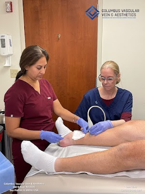
What to expect during a venous duplex exam
- Short intake and symptom review, then exposure of the leg from groin to ankle Warm gel and a linear probe to scan superficial and deep veins, first in the groin, then down Standing or tilted positioning to let gravity challenge valves, with calf squeeze and release Color and Doppler recordings to measure reflux duration and confirm flow direction Marking of target veins on the skin and a clear briefing on findings and options
The entire visit, including discussion, usually takes 30 to 60 minutes. There is no radiation. There is no need to fast. Hydrate and avoid applying heavy lotions that can interfere with the gel.
Screening quality and why some clinics stand out
Accuracy depends on protocol, training, and equipment. High frequency linear transducers between 5 and 12 MHz visualize superficial trunks and tributaries. Color gain and scale must be set so flow fills the lumen without oversaturation. Reflux testing without proper provocation undercounts disease. A clinic that performs scans sitting or supine for convenience will miss pathology. Ask who performs the scan, what credentials they hold, and whether your mapping includes named junctions, measured reflux times, and vein diameters. How accurate are vein clinic screenings? In experienced centers that standardize their approach, very. In settings where the scan is rushed or delegated without oversight, less so.
Ultrasound at the bedside of symptoms patients often misread
- Itchy varicose veins are not just dry skin. Ultrasound often shows chronic superficial reflux feeding inflamed tributaries. Clinics treat it by closing the source and, if needed, injecting the surface veins. Pruritus usually calms as pressure drops. Night leg cramps can reflect muscle fatigue, but when they pair with evening swelling or restlessness, duplex often reveals reflux. Vein clinics can help with leg cramps at night by addressing the reflux, encouraging hydration, and guiding magnesium only when appropriate. Suddenly darker veins on the legs raise concern. We scan to exclude clot in a superficial varix and to assess for new reflux. Sometimes heat or recent travel explains the surge. Bulging veins in summer heat make sense on ultrasound. Vasodilation increases the reservoir, and the failing valve shows longer reflux. Cooling, elevation, and compression socks can blunt the seasonal swing.
Travel, flying, and the scan that protects you
Long flights combine immobility, dehydration, and lower cabin humidity. They do not permanently worsen varicose veins, but they raise the risk of swelling and, in predisposed individuals, clots. A pre travel ultrasound is not standard for everyone, but in a patient with unilateral swelling, recent calf pain, or a history of clot, it is prudent. For frequent travelers, we tailor tips based on the map. If the deep system is pristine and the saphenous trunk shows mild reflux, compression during travel and simple ankle pumps suffice. If a residual clot lingers in a calf vein, we change the approach and sometimes postpone elective procedures.

Compression socks and their real role, revealed by imaging
Do compression socks really prevent vein disease? They do not cure valve failure, but ultrasound shows what they achieve. By increasing external pressure, they reduce vein caliber and limit reflux duration, particularly in smaller tributaries and perforators. Patients often notice lighter legs by evening and fewer cramps. After treatment, compression supports the closed segment and minimizes bruising. The degree of benefit varies. Ultrasound helps set realistic expectations and decide the right strength and length.
Diet, hydration, and small habits that nudge your ultrasound in the right direction
Vein walls contain collagen and elastin. A diet with adequate protein, vitamin C, and flavonoids supports the matrix that resists dilation. No supplement can reverse a failing valve, but in my practice, patients who prioritize hydration, maintain a healthy weight, and walk daily recover faster and report fewer late day symptoms. Obesity complicates treatment by increasing abdominal pressure and making access more challenging. Rapid weight loss can temporarily make superficial veins look more prominent as subcutaneous fat thins. Ultrasound helps separate cosmetic visibility from true hemodynamic change.
Why ultrasound is central to outcomes, not an add on
Technology improves vein treatment outcomes by guiding every critical decision. We avoid the saphenous nerve by knowing where the vein travels alongside it. We prevent skin burns by measuring depth and tumescent anesthesia spread in real time. We detect and treat tributaries that would otherwise re pressurize a treated segment. Closure rates for thermal ablation exceed 90 percent at 1 year when ultrasound is used meticulously before, during, and after the procedure. Lower rates almost always trace back to poor mapping, inadequate energy delivery, or untreated feeders. Ultrasound helps clinics personalize vein treatment plans and explains why customized treatment matters for vein health.
The follow up scans that safeguard the result
Patients often ask how soon you see results from vein treatments. Symptoms such as heaviness often improve within days. Visible changes unfold over weeks as tributaries shrink. We schedule a follow up ultrasound within about a week to confirm closure and exclude rare complications like endothermal heat induced thrombosis. Additional scans at 3 months and, if needed, 6 to 12 months track progress. These brief visits let us fine tune recovery, address new perforators early, and calibrate compression. They also catch the few cases where veins reappear after treatment, giving us a chance to prevent recurrence after vein procedures before it becomes a bigger issue.
Sleep, stress, and the subtle effects on the venous map
Poor sleep and high stress raise cortisol and sympathetic tone, which can affect perception of leg discomfort and amplify cramping. While these do not change valve function directly, patients who improve sleep hygiene often tolerate compression better and recover more smoothly. After vein treatment, sleeping with the leg slightly elevated for a few nights helps. Side sleeping with a pillow between the knees reduces hip rotation that can tug on the groin area after a great saphenous closure. These are small adjustments, but they make the first week easier.
Common missteps and how ultrasound prevents them
Skipping ultrasound leads to three predictable mistakes. First, treating only what you see at the skin and ignoring the feeder vein. Second, ablating a segment without confirming adequate tumescent anesthesia depth and spread, which increases pain and bruising. Third, overlooking deep venous obstruction that limits symptom relief from superficial treatment alone. With a complete ultrasound, we avoid these traps. We also prevent rare but serious problems. For example, if a small saphenous vein drains high into a muscular branch rather than the popliteal vein, knowing that anatomy in advance prevents misplacement of thermal energy close to the tibial nerve.
A brief case that captures the point
A 47 year old pediatric nurse came in with evening ankle swelling, tightness, and a few new spider veins. She stood on the ward most of the day and had started wearing inexpensive compression socks. Someone suggested injections. Her duplex mapping showed 0.8 second reflux in the great saphenous vein from mid thigh to mid calf, two incompetent perforators near the medial ankle, and a clean deep system. We closed the refluxing trunk with radiofrequency, injected the perforators under ultrasound, and asked her to hydrate and walk after the procedure. At 1 week, ultrasound confirmed closure and no heat induced thrombus. At 3 months, her swelling had resolved, the spiders had faded by half, and she no longer had to peel off tight socks at night. Without ultrasound, we might have chased the surface veins and called it a day, only to see them return.
What happens if vein disease goes untreated
Mild reflux can remain stable for years, but in many, pressure slowly damages the ankle skin. Hemosiderin staining darkens the lower leg, eczema flares, and ulcers can open with minor trauma. Quality of life degrades long before those endpoints. Ultrasound does not obligate you to treatment, but it informs your choices. It shows whether daily walking and compression are enough or whether a focused intervention would spare your skin and stamina.
Choosing where to have your scan and treatment
A trustworthy vein clinic feels different at the first visit. The team explains what reflux is in plain terms. They scan you standing when safe. They time reflux, measure vein diameters, and draw a map you can understand. They discuss options, including doing nothing beyond lifestyle steps when that is reasonable. They welcome questions patients forget to ask vein specialists, such as how they track progress after treatment, how they manage expectations, and realistic timelines for vein treatment results. They tell you how long vein procedures actually take and what recovery really feels like. They talk openly about recurrence risks and long term maintenance strategies.
The quiet power of a simple tool
An ultrasound exam does not look glamorous. There is no dramatic monitor beeping or complex console to impress anyone. What it delivers, though, is clarity. It ties your symptoms to a visible, measurable flow problem. It separates superficial from deep disease. It warns us about nerves, tributaries, and tortuous paths before a catheter ever touches your skin. It personalizes your plan and guards your result. For a field once driven by educated guesses and big incisions, that is progress you can feel every time you take the stairs without heaviness.
If your legs tire early, if your veins itch, if your ankles swell by evening, do not rely on mirrors and hunches. Ask for a proper venous duplex ultrasound. The probe, the gel, and twenty minutes of mapping may be the most important steps you take Des Plaines, IL vein clinic toward better circulation and lighter legs.